|
Move mouse coursor or your finger upon an image
and you can see the image without labells
|
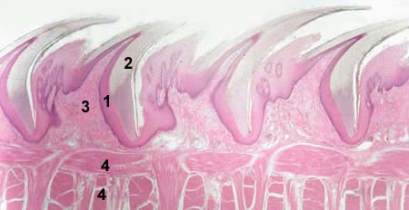
FILIFORM PAPILLAE OF THE TONGUE
Stained with haematoxylin and eosin
1 - epithelium covering papilla
(stratified squamous keratinizing)
2 - keratinized layer of the epithelium
3 - core of the papilla (lamina propria
of the mucosa of dorsal surface of the tongue)
2 - tongue muscles
|
|
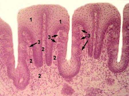
LEAF SHAPED PAPILLAE OF THE TONGUE
Stained with haematoxylin and eosin
1 - epithelium covering papilla
(stratified squamous nonkeratinizing)
2 - core of the papilla (lamina propria
of the mucosa of dorsal surface of the tongue)
3 - taste buds
|
|
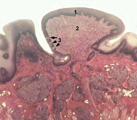
CIRCUMVALLATE PAPILLAE OF THE TONGUE
Stained with haematoxylin and eosin
1 - epithelium covering papilla
(stratified squamous nonkeratinizing)
2 - core of the papilla (lamina propria
of the mucosa of dorsal surface of the tongue)
3 - taste buds
|
|
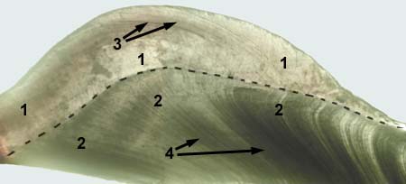
TOOTH
Nonstained
1 - enamel
2 - dentine
3 - Retcius lines
4 - dentine tubules
|
|
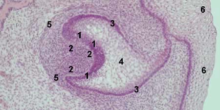
TOOTH DEVELOPMENT - ENAMEL ORGAN
Stained with haematoxylin and eosin
1, 3, 4 - enamel organ
1 - internal cells of the enamel organ
(these cells will be ameloblsats)
2 - dental papilla
3 - external cells of the enamel organ
4 - cells forming the main bulk of
the enamel organ (stellate reticulum)
5 - dental follicle
6 - epithelium of oral cavity
|
|
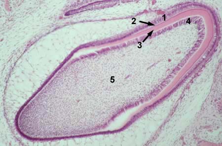
TOOTH DEVELOPMENT - FORMATION OF DENTAL TISSUES
Stained with haematoxylin and eosin
1 - ameloblasts (former external cells of the enamel organ)
2 - enamel
3 - dentine (predentine)
4 - odontoblasts (cells which covered the top of dental papilla)
5 - dental pulp (former dental papilla)
|
|
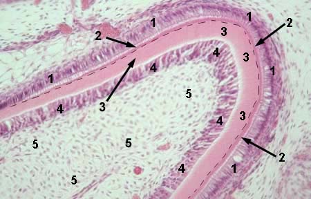
TOOTH DEVELOPMENT - FORMATION OF DENTAL TISSUES
Stained with haematoxylin and eosin
1 - ameloblasts
2 - enamel
3 - dentine (predentine)
4 - odontoblasts
5 - dental pulp
border between enamel and dentine
is marked with dot line
|
|