|
|
Move mouse coursor or your finger upon an image and you can see the image without labells
|
|
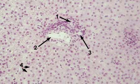
LIVER Stained with haematoxylin and eosin 1 - hepatic artery
|
|
|
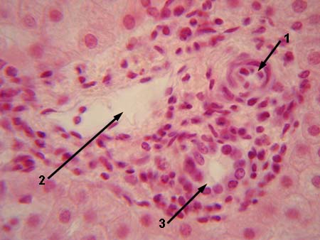
LIVER Stained with haematoxylin and eosin 1 - hepatic artery
|
|
|
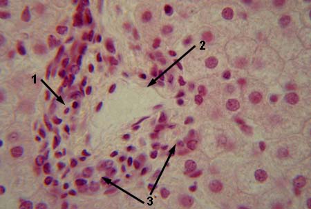
LIVER Stained with haematoxylin and eosin 1 - hepatic artery
|
|
|
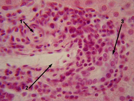
LIVER Stained with haematoxylin and eosin 1 - hepatic artery
|
|
|
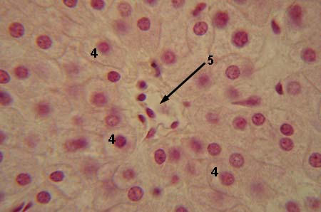
LIVER Stained with haematoxylin and eosin 4 - hepatocytes
|
|
|
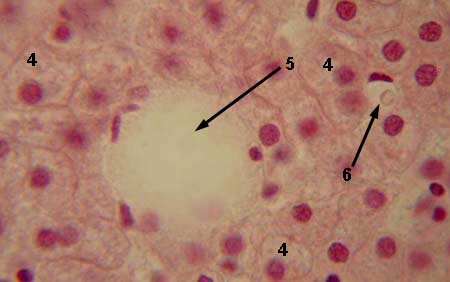
LIVER Stained with haematoxylin and eosin 4 - hepatocytes
|
|
|
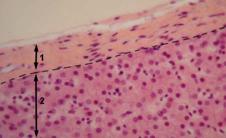
LIVER Stained with haematoxylin and eosin 1 - capsule
|
|
|
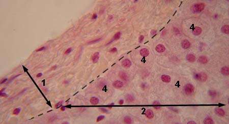
LIVER Stained with haematoxylin and eosin 1 - capsule
|
|
|
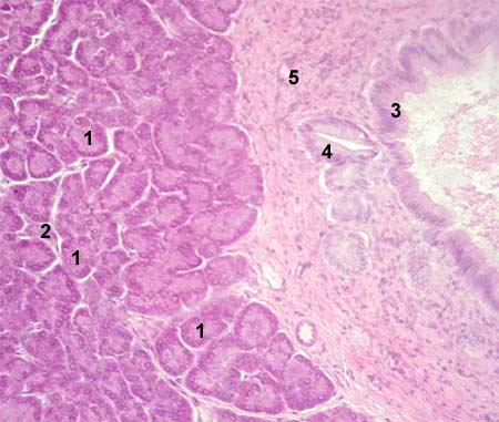
PANCREAS Stained with haematoxylin and eosin 1 - glandular acinus
|
|
|
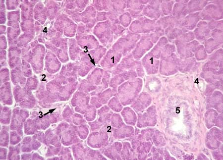
PANCREAS Stained with haematoxylin and eosin 1 - acinus
|
|
|
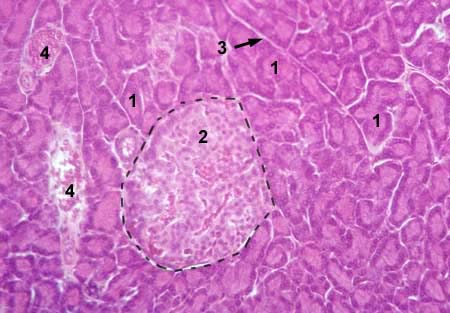
PANCREAS Stained with haematoxylin and eosin 1 - acinus
|
|
|
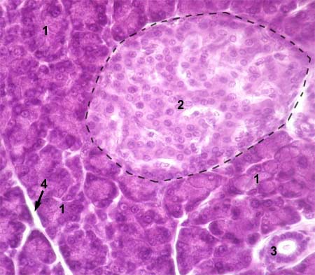
PANCREAS Stained with haematoxylin and eosin 1 - acinus
|
|