|
|
Move mouse coursor or your finger upon an image and you can see the image without labells
|
|
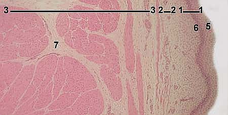
URINARY BLADDER Stained with haematoxylin and eosin 1 - tunica mucosa
|
|
|
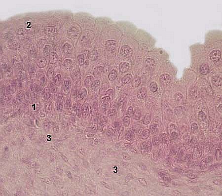
URINARY BLADDER transitional epithelium of the mucosa Stained with haematoxylin and eosin 1 - basal layer of the epithelium
|
|
|
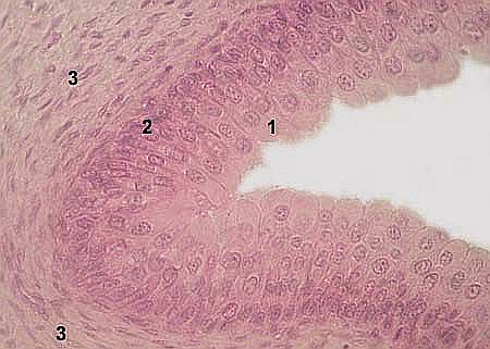
URINARY BLADDER transitional epithelium of the mucosa Stained with haematoxylin and eosin 1 - basal layer of the epithelium
|
|
|
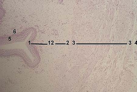
URETER Stained with haematoxylin and eosin 1 - tunica mucosa
|
|
|
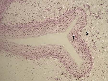
URETER transitional epithelium of the mucosa Stained with haematoxylin and eosin 1 - transitional epithelium
|
|