|
Move mouse coursor or your finger upon an image
and you can see the image without labells
|
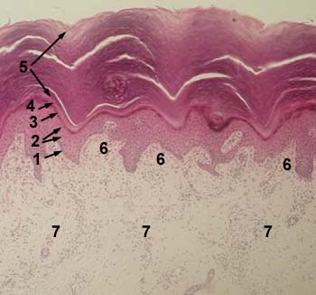
SKIN OF THE FINGER
Stained with haematoxylin and eosin
1 - basal layer of epidermis
2 - prickle cell layer (stratum spinosum) of epidermis
3 - granular layer of epidermis
4 - lucidar layer of epidermis
5 - cornified layer of epidermis
6 - papillary layer of dermis
7 - reticular layer of dremis
|
|
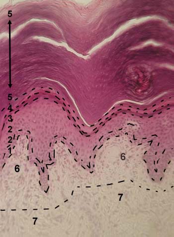
SKIN OF THE FINGER
Stained with haematoxylin and eosin
1 - basal layer of epidermis
2 - prickle cell layer (stratum spinosum) of epidermis
3 - granular layer of epidermis
4 - lucidar layer of epidermis
5 - cornified layer of epidermis
6 - papillary layer of dermis
7 - reticular layer of dremis
|
|
SKIN OF THE FINGER
Stained with haematoxylin and eosin
1 - basal layer of epidermis
2 - prickle cell layer (stratum spinosum) of epidermis
3 - granular layer of epidermis
4 - lucidar layer of epidermis
5 - cornified layer of epidermis
6 - papillary layer of dermis
7 - reticular layer of dremis
|

|
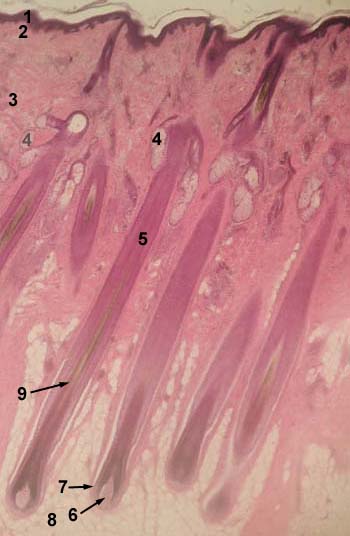
HAIR SKIN
Stained with haematoxylin and eosin
1 - epidermis
2 - papillary layer of dermis
3 - reticular layer of dremis
4 - sebaceous gland
5 - hair follicle
6 - dermal papilla
7 - hair bulb
8 - hypodermis
9 - hair root
|
|
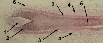
HAIR FOLLICLE
Stained with haematoxylin and eosin
1 - dermal papilla
2 - hair bulb
3 - inner root sheath
4 - outer root sheath
5 - hair root
6 - connective tissue sheath
|
|