| CONTENTS |
HISTOLOGY IMAGES
|
WEB HISTOLOGY TEXTBOOK |
SENSE ORGANS
|
|
Move mouse coursor or your finger upon an image and you can see the image without labells
|
|
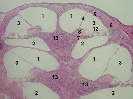
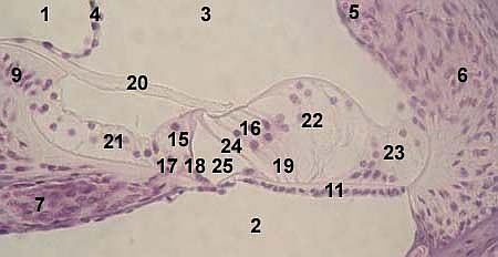
ORGAN OF CORTI (organ of hear) Stained with haematoxylin and eosin
1 - scala vestibuli
15 - inner sensory (hair) cells
|
|
|
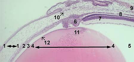
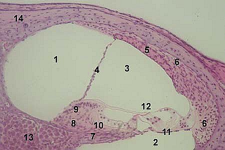
EYE Stained with haematoxylin and eosin 1 - cornea
|
|
|
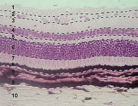
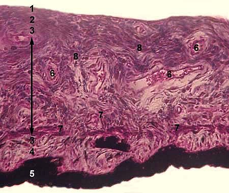
EYE: RETINA, CHOROID Stained with haematoxylin and eosin 1 - 8 - retina
|
|
|
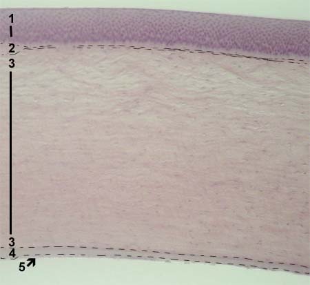
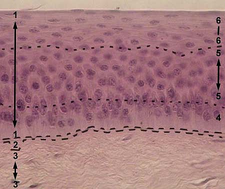
EYE: CORNEA Stained with haematoxylin and eosin 1 - anterior epithelium (stratified
|
|
|
EYE: CORNEA Stained with haematoxylin and eosin anterior epithelium (stratified squamous epithelium) 2 - anterior basement (Bowman's) membrane 3 - substantia propria 4 - basal layer of the epithelium 5 - intermediate layer of the epithelium 6 - superficial layer of the epithelium
|
|
|
EYE: IRIS Stained with haematoxylin and eosin 1 - anterior epithelium
|
|
|
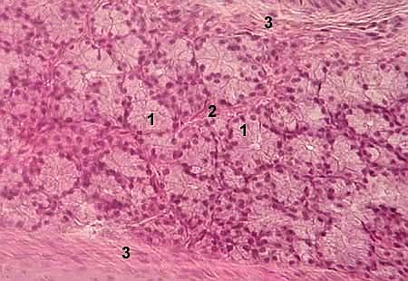
LACRIMAL GLAND Stained with haematoxylin and eosin 1 - secretory (end) unit
|
|
|
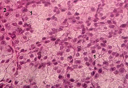
LACRIMAL GLAND Stained with haematoxylin and eosin 1 - secretory (end) unit
|
|
|
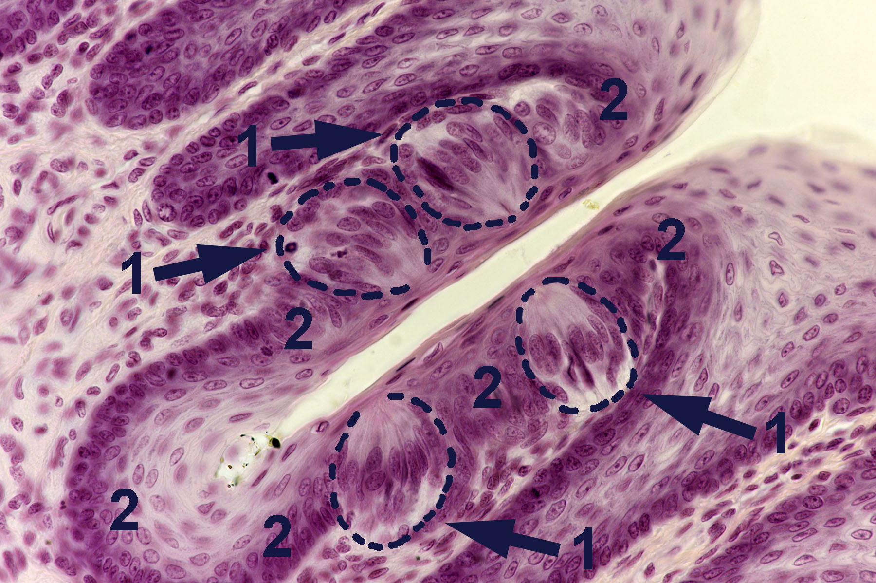
TASTE BUDS Stained with haematoxylin and eosin 1 - taste buds
|
|
|
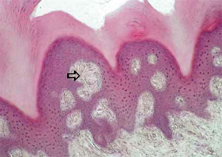
MEISSNER'S CORPUSCLE in the dermis of the skin of the fingertip Stained with haematoxylin and eosin Meissner's corpuscle (oval in shape)
|
|
|
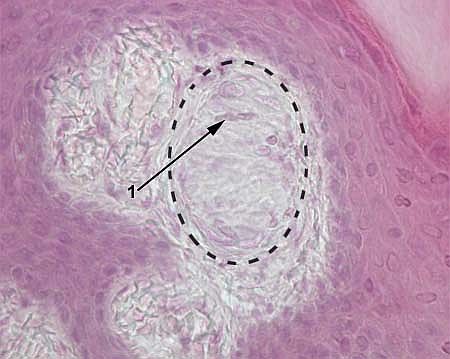
MEISSNER'S CORPUSCLE in the skin of the fingertip Stained with haematoxylin and eosin Meissner's corpuscle encircled
with dotted line
|
|
|
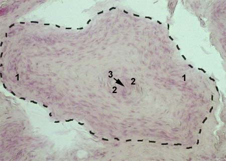
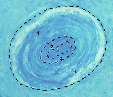
PACINIAN CORPUSCLE Stained with haematoxylin and eosin Pacinian corpuscle encircled
|
|
|
HERBST AND GRANDRY SENSORY CORPUSCLE Stained with haematoxylin and eosin Herbst and Grandry corpuslcle encircled
|
|
© histol@mail.ru