| CONTENTS |
HISTOLOGY IMAGES
|
WEB HISTOLOGY TEXTBOOK |
NERVOUS TISSUE
|
|
Move mouse coursor or your finger upon an image and you can see the image without labells
|
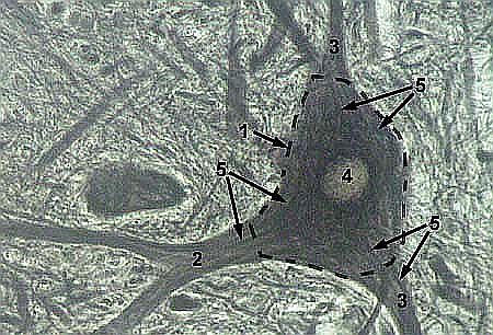
| NEURONE Impregnated with aurum chloride 1 - body of the nervous cell (pericaryon)
|
 |
| NEURONE Impregnated with silver nitrate 1 - body of the nervous cell (pericaryon)
|
 |
| NEURONE: neurofibrills Impregnated with silver nitrate 1 - body of the nervous cell (pericaryon)
|
 |
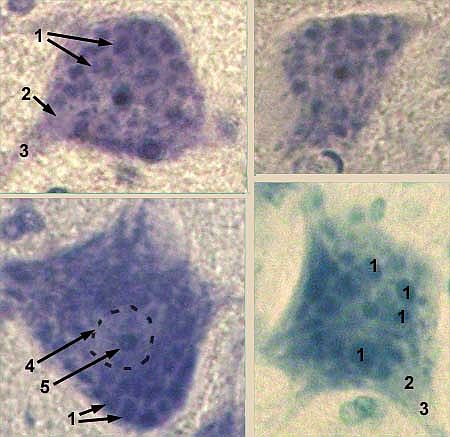
| NEURONE: Nissl substance Stained with toluidine blue by Nissl method 1 - ribosomes form clusters which are stained |
 |
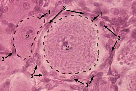
| NEURONE Stained with haematoxylin and eosin 1 - body of the nervous cell (pericaryon)
|
 |
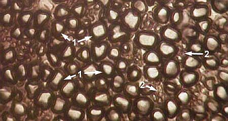
| PERIPHERAL NERVE (transverse section) Stained with osmium oxide 1 - myelinated nerve fibers (myelin sheath |
 |
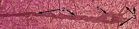
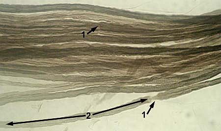
| MYELINATED NERVE FIBRES Node of Ranvier is indicated by arrow Stained with osmium oxide 1 - node of Ranvier
|
 |
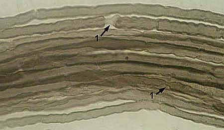
| MYELINATED NERVE FIBRES Stained with osmium oxide 1 - node of Ranvier
|
 |
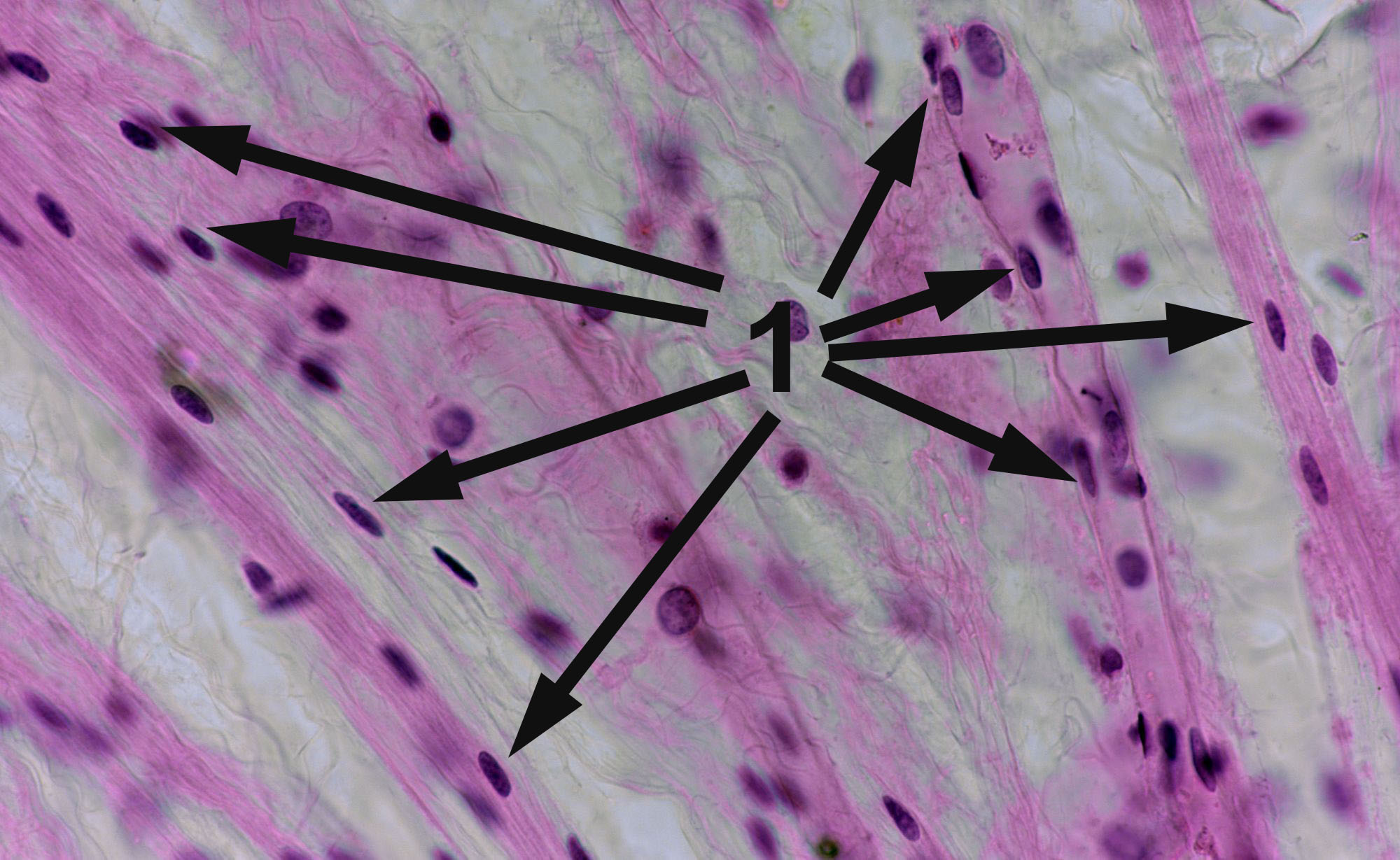
| NONMYELINATED NERVE FIBRES Stained with haematoxylin and eosin 1 - nuclei of Schwann cells
|
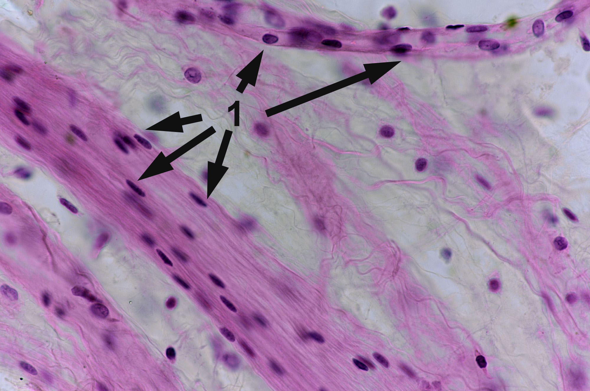 |
| NONMYELINATED NERVE FIBRES Stained with haematoxylin and eosin 1 - nuclei of Schwann cells
|
 |
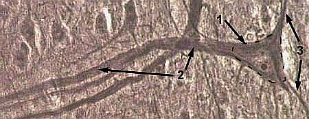
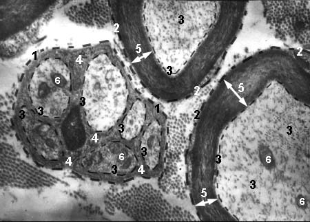
| MYELINATED AND NONMYELINATED NERVE FIBRES Electron microscopy image
1 - nonmyelinaled nerve fibre |
 |
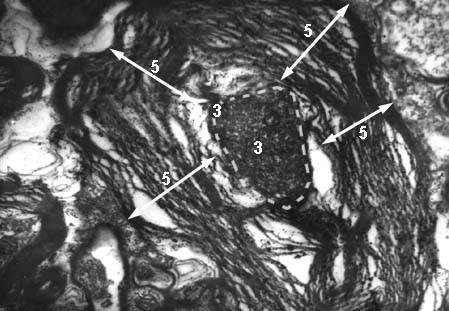
| PATHOLOGICAL MYELINATED NERVE FIBRE Electron microscopy image
3 - wrinkled axon |
 |
© histol@mail.ru