| CONTENTS |
HISTOLOGY IMAGES
|
WEB HISTOLOGY TEXTBOOK |
IMMUNE SYSTEM
|
|
Move mouse coursor or your finger upon an image and you can see the image without labells
|
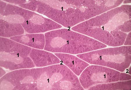
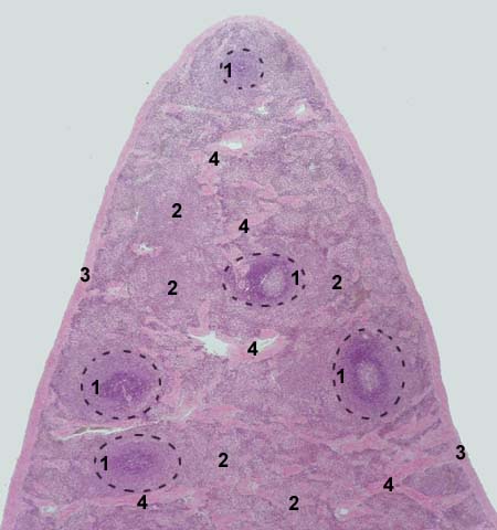
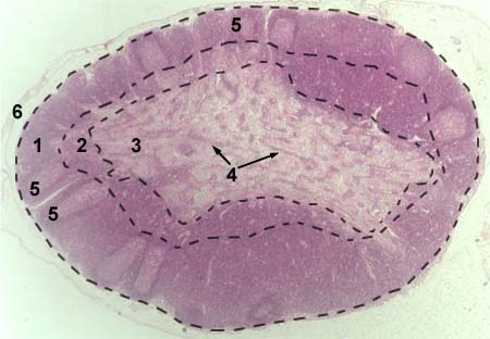
| THYMUS
Stained with haematoxylin and eosin 1 - lobules
|
 |
| THYMUS
Stained with haematoxylin and eosin 1 - lobules
|
 |
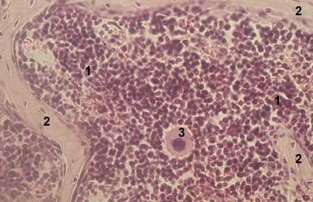
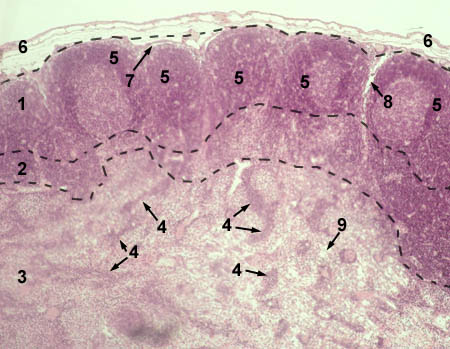
| THYMUS (lobule)
Stained with haematoxylin and eosin 1 - cortex
|
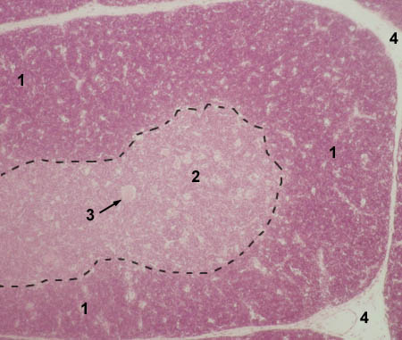 |
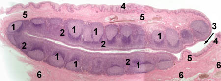
| THYMUS (Hassal's corpuscle)
Stained with haematoxylin and eosin 1 - Hasal's corpuscle
|
 |
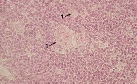
| THYMUS (Hassal's corpuscle)
Stained with haematoxylin and eosin 1 - Hassal's corpuscle
|
 |
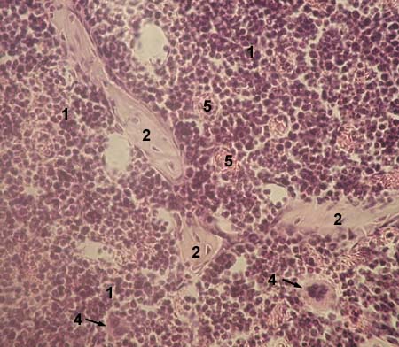
| RED BONE MARROW
Stained with haematoxylin and eosin 1 - parenchyma of red marrow (haemopoietic cells and reticular stroma)
|
 |
| RED BONE MARROW
Stained with haematoxylin and eosin 1 - parenchyma of red marrow (haemopoietic cells and reticular stroma)
|
 |
| SPLEEN
Stained with haematoxylin and eosin 1 - lymphoid follicle (white pulp)
|
 |
| SPLEEN (follicle)
Stained with haematoxylin and eosin lymphoid follicle is circled with
|
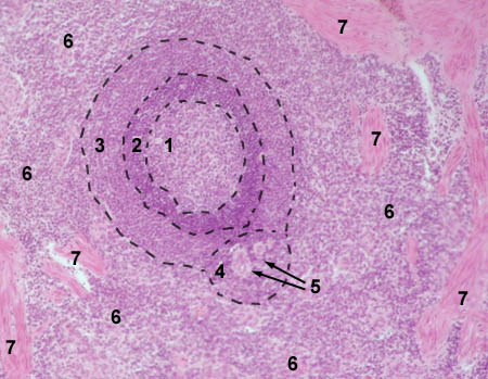 |
| SPLEEN (follicle)
Stained with haematoxylin and eosin lymphoid follicle is circled with
|
 |
| LYMPH NODE
Stained with haematoxylin and eosin 1 - cortex
|
 |
| LYMPH NODE
Stained with haematoxylin and eosin 1 - cortex
|
 |
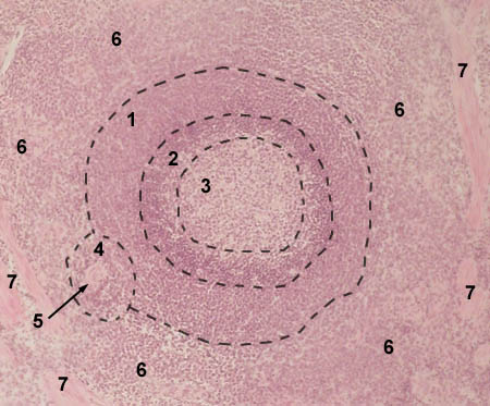
| PALATINE TONSIL
Stained with haematoxylin and eosin 1 - lymphoid follicle
|
 |
© histol@mail.ru