| CONTENTS |
HISTOLOGY IMAGES
|
WEB HISTOLOGY TEXTBOOK |
EPITHELIAL TISSUE
|
|
Move mouse coursor or your finger upon an image and you can see the image without labells
|
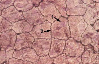
| MESOTHELIUM (SIMPLE SQUAMOUS EPITHELIUM) view from surface Stained with silver nitrate 1 - nucleus of cell |
 |
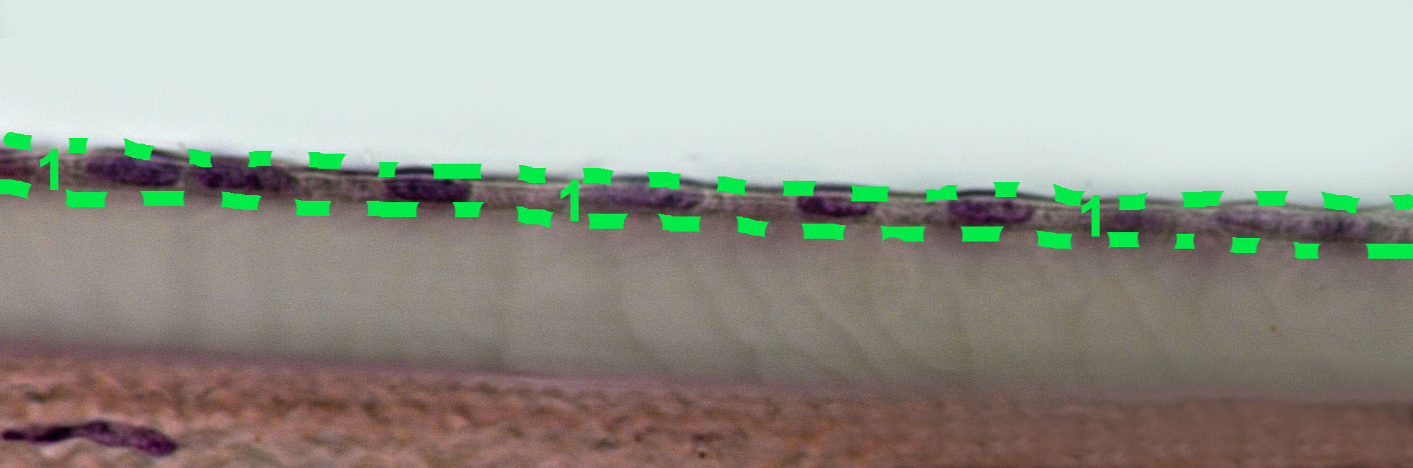
| SIMPLE SQUAMOUS EPITHELIUM Stained with haematoxylin and eosin Nuclei of the epithelial cells are shown with an arrow
|
 |
| SIMPLE CUBOIDAL EPITHELIUM Stained with haematoxylin and eosin
|
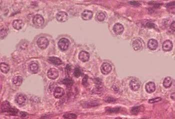 |
| SIMPLE CUBOIDAL EPITHELIUM Stained with haematoxylin and eosin |
 |
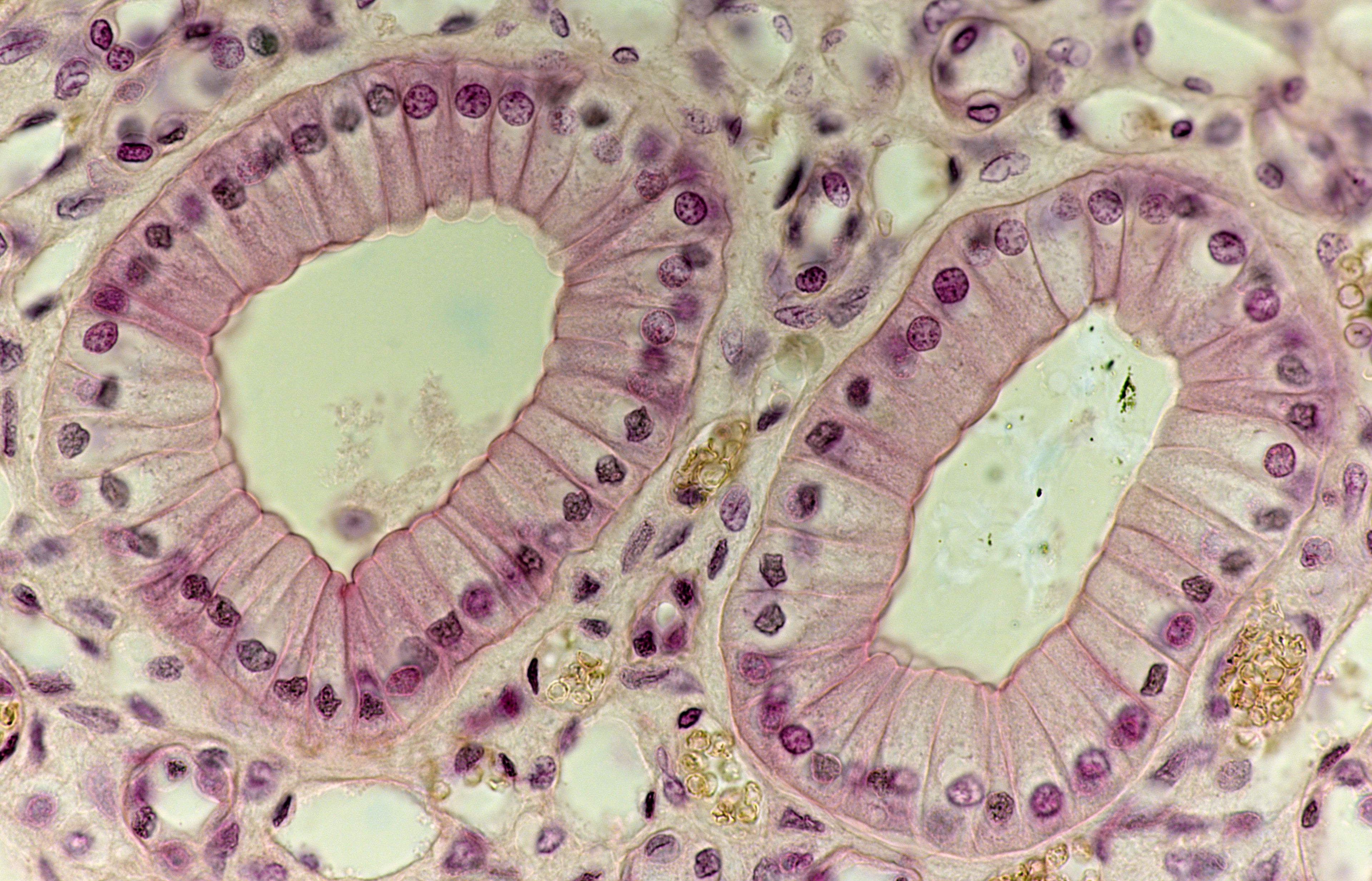
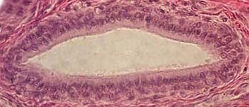
| SIMPLE COLUMNAL EPITHELIUM Stained with haematoxylin and eosin |
 |
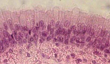
| SIMPLE PSEUDOSTRATIFIED COLUMNAR EPITHELIUM Stained with haematoxylin and eosin |
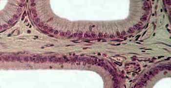 |
| SIMPLE PSEUDOSTRATIFIED COLUMNAR CILIATED EPITHELIUM Stained with haematoxylin and eosin |
 |
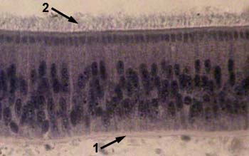
| SIMPLE PSEUDOSTRATIFIED COLUMNAR CILIATED EPITHELIUM Stained with iron haematoxylin
1 - basal membrane |
 |
| STRATIFIED (TWO-LAYER) COLUMNAR EPITHELIUM Stained with haematoxylin and eosin |
 |
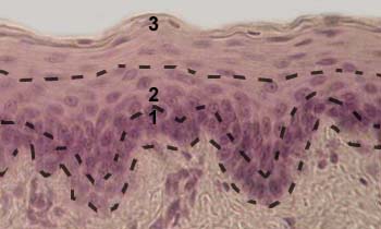
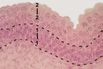
| STRATIFIED SQUAMOUS NONKERATINISING EPITHELIUM Stained with haematoxylin and eosin
1 - basal layer |
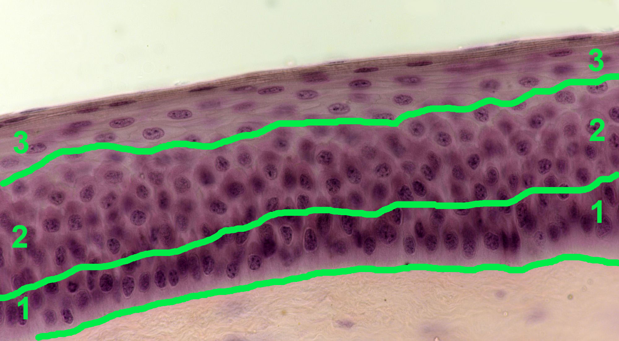 |
| STRATIFIED SQUAMOUS NONKERATINISING EPITHELIUM Stained with haematoxylin and eosin
1 - basal layer |
 |
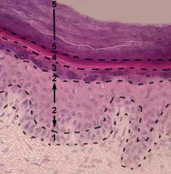
| STRATIFIED SQUAMOUS KERATINISING EPITHELIUM (EPIDERMIS) Stained with haematoxylin and eosin
1 - basal layer |
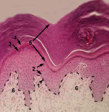 |
| STRATIFIED SQUAMOUS KERATINISING EPITHELIUM (EPIDERMIS) Stained with haematoxylin and eosin
1 - basal layer |
 |
| TRANSITIONAL EPITHELIUM (UROTHELIUM) Stained with haematoxylin and eosin
1 - basal layer |
 |
© histol@mail.ru