|
|
Move mouse coursor or your finger upon an image and you can see the image without labells
|
|
THYROID GLAND
Stained with haematoxylin and eosin 1 - thyroid follicle
|

|
|
THYROID GLAND
Stained with haematoxylin and eosin 1 - wall of the thyroid follicle, built from thyrocytes
|

|
|
THYROID GLAND
Stained with haematoxylin and eosin 1 - wall of the thyroid follicle, built from thyrocytes
|

|
|
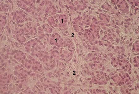
PARATHYROID GLAND
Stained with haematoxylin and eosin 1 - parathyrocytes
|
|
|
ADRENAL GLAND (SUPRARENAL GLAND) Stained with haematoxylin and eosin 1 - outer capsule of the adrenal gland
|

|
|
ADRENAL GLAND (SUPRARENAL GLAND) zona glomerulosa of the cortex Stained with iron haematoxylin 1 - endocrine cells of the zona glomerulosa
|

|
|
ADRENAL GLAND (SUPRARENAL GLAND) zona fasciculata of the cortex Stained with iron haematoxylin 1 - endocrine cells of the zona fasciculata
|

|
|
ADRENAL GLAND (SUPRARENAL GLAND) zona reticularis of the cortex Stained with iron haematoxylin 1 - endocrine cells of the zona reticularis |

|