| CONTENTS |
HISTOLOGY IMAGES
|
WEB HISTOLOGY TEXTBOOK |
CYTOLOGY
|
|
Move mouse coursor or your finger upon an image and you can see the image without labells
|
|
GLYCOGEN IN LIVER CELLS Stained with carmin, nuclei are stained with haematoxylin
1 - glycogen (red or magenta staining)
|
|
|
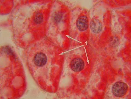
GLYCOGEN IN LIVER CELLS
Stained with carmin, nuclei are stained with haematoxylin 1 - glycogen (red or magenta staining)
|
|
|
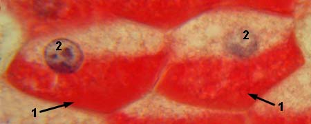
GLYCOGEN IN LIVER CELLS
Stained with carmin, nuclei are stained with haematoxylin
1 - glycogen (red or magenta staining)
|
|
|
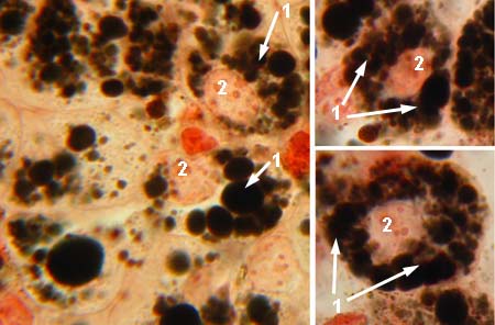
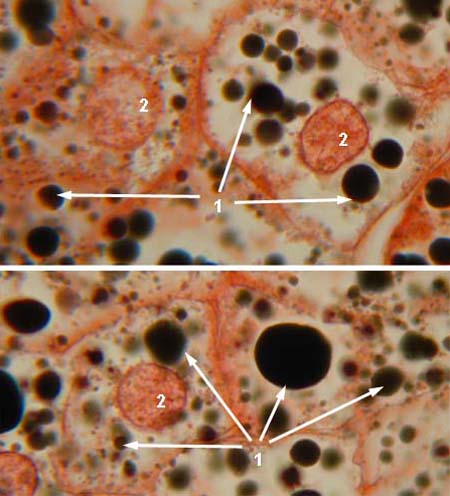
LIPIDS IN LIVER CELLS
Stained with osmium, nuclei are stained with safranin
1 - lipid droplets (black)
|
|
|
LIPIDS IN LIVER CELLS
Stained with osmium, nuclei are stained with safranin
1 - lipid droplets (black)
|
|
|
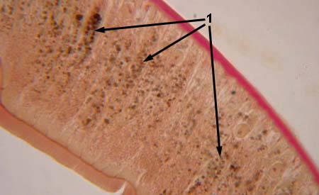
GOLGI BODY
Stained with silver nitrate
1 - Golgi body (dark curls or ringlets)
|
|
|
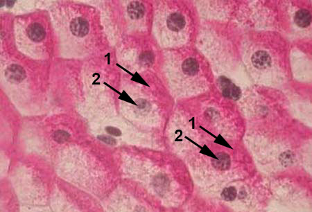
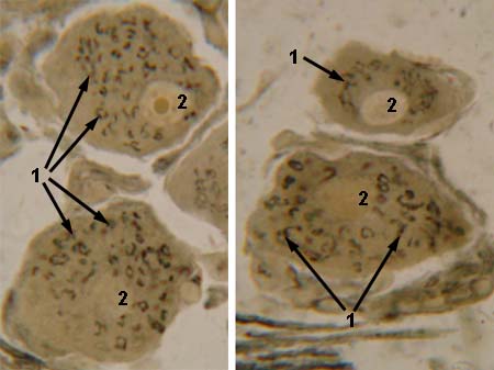
MITOCHONDRIA IN EPITHELIAL CELLS
Stained with silver nitrate, nuclei are stained with safranin
1 - mitochondria (dark dots)
|

|
|
MITOCHONDRIA IN EPITHELIAL CELLS
Stained with silver nitrate, nuclei are stained with safranin
1 - mitochondria (dark dots)
|
|
|
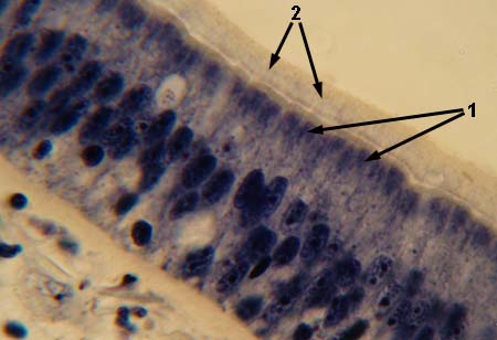
CILIA (IN CILIATED EPITHELIAL CELLS)
Stained with iron haematoxylin
1 - basal bodies of cilies
|
|
|
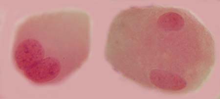
AMITOSIS
Stained with haematoxylin and eosin cells which divide by amitosis
|
|
|
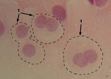
AMITOSIS
Stained with haematoxylin and eosin cells which divide by amitosis
|
|
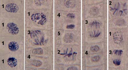
| MITOSIS IN PLANT CELLS
Stained with iron haematoxylin
1 - prophase
|
|
|
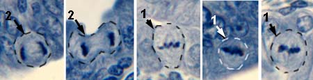
MITOSIS IN MAMMALIAN CELLS
Stained with iron haematoxylin
1 - metaphase |
|
© histol@mail.ru